Considerable advancements in nanotechnology over the last couple of decades have both challenged and pushed forward the development of enhanced biosensor techniques beyond the conventional ones. Quantum dots (QDs) have received far greater attention as an emerging technology of the future due to their exceptional benefits. In this article, we highlight unique advantages of quantum dots, recent progress made in their development, and bioconjugation of quantum dots.
Methods for bioconjugation of quantum dots include covalent binding, noncovalent binding, or direct coupling of ligands to coat the surface of QDs. Quantum dots can be attached to DNA, Proteins, and numerous other biomolecules.
Quantum dots (QDs) were first theorized in the 1970s. However, it was not until 1981 that the first semiconductor nanocrystal was successfully synthesized by a Russian physicist, Alexei Ekimov. Since then, QDs have surmounted various fundamental scientific challenges and augmented the imagination of researchers due to their versatility, sensitivity and quantitative capabilities.
What are QuantumDots?
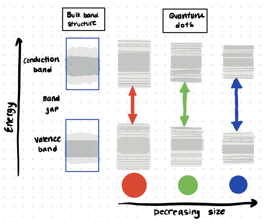
Quantum dots (QDs) are a class of semiconductor nanocrystals which exhibit size-dependent optoelectronic properties. They emit intense luminescence and the color of the luminescence can be varied by changing the size of the quantum dots.
They present intense luminescence with a wide range of emission bandwidths due to their shape, form, and the quantum confinement effect in three dimensions (Kargozar et al., 2020; Zhao and Zeng, 2015; Reshma and Mohanan, 2018). An alternative method for labeling biomolecules which was utilized in the past was radiopharmaceutical chemistry. Attachment of radioisotopes gives similar levels of detection as quantum dots, but, as you can imagine, it’s not as easy to get a radiolabeling lab set up and scale up is challenging.
Types of Quantum Dots
One of the most attractive properties of QDs is the fact that their emission spectra can be finely tuned, merely by varying their shape, size, and material composition (Cotta, 2020). With current advances in bioconjugation chemistry and the availability of different components available for functionalization, there has been increasing interest in combining QDs with other materials to create multimodal nanocomposites.
Different types of quantum dots include colloidal quantum dots which are used for biosensing, magnetic quantum dots which enable magnetic cell separation, and fluorescent quantum dots which can be utilized for MRI.
Colloidal Quantum Dots
Colloidal QDs (CQDs) are synthesized by heating precursor solutions at high temperature—causing them to decompose and form nucleated monomers to generate nanocrystals. To facilitate their dispersion in solvents and prevent agglomeration, the reaction solution is typically employed with surfactants/ligands that can coat the CQDs.
Colloidal quantum dots, especially those made of CdS, CdSe, CdTe, PbS, and PbSe, are the most widely used form of QDs due to their efficient properties and their ability to self-assemble into an ordered superlattice (Benehkohal et al., 2021).
So far, CQDs have found the widest range of applications in bioanalytics and biolabeling. However, research around the CQDs for next-generation optoelectronic applications, such as and high-performance QD photovoltaics, lasers, MIR photodetectors, single-photon emitters, etc. are still going strong (Liu et al., 2021).
Magnetic Quantum Dots
The combination of gold, MNPs, and ions with QDs represent a new and captivating class of nanocomposite with a considerable number of applications in bioimaging, diagnosis and treatment. Magnetic QDs (MQDs) generally consist of a hydrophobic magnetic nanoparticle core surrounded by a shell of QDs and encapsulated in a reverse micelle-based coating.
Magnetic quantum dots possess excellent superparamagnetic and intense fluorescent properties—allowing them to easily target, separate, and be used to visualize any biomolecule simply by engineering the magnetic fields. Today, MQDs have been utilized for a variety of biochemical applications, including magnetic-activated cell separations, targeted drug delivery, and as contrast agents for MRI.
For more on magnetic nanoparticles (MNPs) and their applications, read our article, magnetic nanoparticle bioconjugation.
Fluorescent Quantum Dots
Apart from acting as fluorophores, QDs also show remarkable activity as quenchers via either electron transfer or FRET.
Fluorescent QDs, especially the ones based on graphene oxide (GQDs) have found extensive applications as biosensors and chemi-sensors in medical practice, environment, and agriculture. Graphere quantum dots provide a chemically tunable platform that is inexpensive, non-toxic, photo-stable, water-soluble, biocompatible, and environmentally friendly (Zheng and Wu, 2015; Xu et al., 2014). We talk about an alternative application of biosensing using hydrazine bioconjugation.
Generally, graphene quantum dots are a 3D material generated via hybridization of sp2 and sp3 carbon atoms with graphene, graphite, or other graphite derivatives in a planar (sheet) form. GQDs usually have a layered structure and can be synthesized with either the top-down methods (lateral size of <20 nm), or the bottom-up method (lateral size of <10 nm) (Li et al., 2017; Xu et al., 2014).
Generalized Protocol for Bioconjugation of Quantum Dots
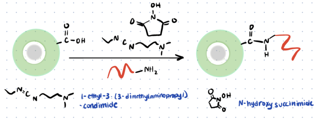
Below we discuss a general protocol for one of the most common QD-DNA bioconjugation strategies via the formation of amide linkages. For a more detailed protocol, read this article. An alternative method involves complexing the DNA with a cationic peptide such as Protamine and then conjugating the QD to the amines and carboxyls on Protamine. Learn more about protamine bioconjugation here.
Trying to attach molecules together? You can explore conjugation kits to help you attach biomolecules together quickly and repeatably here.
Step 1. Functionalization Of Qds
The surface of the QDs must first be functionalized with ligands bearing carboxylic acid functional groups, (MAA, MPA, MHA, DHLA, etc.). In this study, QDs functionalization was conducted with deprotonated 3MPA and TMAH as catalysts in CHCl3 solution (room temperature, 48 h).
Step 2. Removal Of Unbound 3mpa
Alkyl thiols are deactivators of EDC, an important promoter used to couple amine-modified DNA onto the QDs. It is therefore important that excess 3MPA is removed from the QDs solution. These dissolved solutes were removed from the colloid by dialysis against a PBS buffer (10mM, pH 7).
Step 3. Activation Of Carboxylic Acid Group Of Quantum Dots
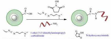
The carboxylic acid group of QDs is activated by reaction with EDC, producing a highly reactive (and labile) O-acylisourea intermediate, which can be immediately mixed with NHS, resulting in QD-NHS. In the case of failure of reaction with NHS, the intermediate hydrolyses and the carboxylic acid on QDs can be regenerated.
Step 4. Formation Of Qd-Dna Bioconjugate
After incubation at room temperature (15 min), amine-modified ssDNA is added to the solution. These NHS-derived QDs are then reacted immediately with amine-DNA to form a stable amide bond between the ligand and DNA (30oC, 30 min)
Step 5. Purification Of Conjugates
The main purification methods for uncoupled DNA are electrophoresis, chromatographic separation, and ultracentrifugation. In this study, the author removed the dissolved solutes by dialysis against a PBS buffer (10mM, pH 7.0) using a cellulose-acetate membrane.
Strategies for Bioconjugation of Quantum Dots
For most of these applications, QDs are first conjugated with biomolecules such as nucleic acids, proteins or signalling molecules.
Strategies for bioconjugation of quantum dots to proteins typically involve covalent/noncovalent linkage. To conjugate quantum dots to fluorophores, a polymer conjugation strategy can be utilized. To attach quantum dots to DNA, it’s possible to do covalent conjugation in aqueous media.
Bioconjugation of Quantum Dots to Proteins
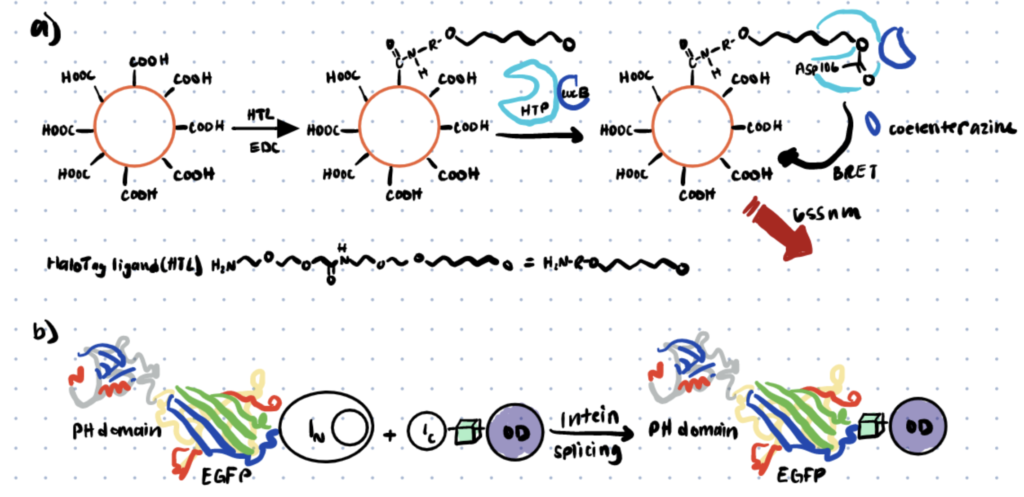
QD-protein bioconjugates have proven to be powerful tools for bioimaging and biosensing. However, the preparation of protein-conjugated QDs is a laborious multi-step process that usually consists of colloidal QDs synthesis, QDs solubilization and biomolecule functionalization.
How you attach your quantum dots to proteins depends on the functional groups present on both molecules. For example, if you have thiols on the surface of your quantum dots, you should consider bioconjugation of maleimides on your proteins to your QDs. If you have carboxyls or NHS esters on your QD surface, then consider lysine conjugation techniques which are simple and well known. If you want site-specific conjugation, consider a rarer amino acid like arginine for conjugation.
Common strategies for conjugation of Quantum dots with proteins include covalent coupling, metal-affinity interaction and avidin-biotin interaction, as summarized by Xing et al.
Bioconjugation of Quantum Dots to DNA
The unique physicochemical/functional properties of DNA have allowed their use as major building blocks in the nano-world. DNA-QD bioconjugates have been successfully prepared mainly by attaching the functional groups of DNA to QDs, which can be done through four different approaches, including:
- EDC coupling chemistry to join amine or carboxyl-functionalized DNA to the cognate groups present on the QD surface. We’ve covered EDC and NHS Ester bioconjugation protocols in more detail here.
- Direct linkage of thiolated DNA to QDs shell surface via dative thiol interactions. We’ve covered thiol-mediated bioconjugation in more detail here.
- Polyhistidine peptide-DNA assembly to QD surfaces via metal-affinity coordination
- Biotinylated DNA attachment to streptavidin-coated QDs.
DNA-QD bioconjugates have become popularly used in bioimaging and detection. More details can be found in this review.
Bioconjugation of Quantum Dots to Fluorophores
QDs have been used as the energy ‘donor’ for the FRET pairs. There are several examples where QDs perform as FRET acceptors, by coupling them with polymers that can act as energy donors, such as PVK, PANI, TPAPAM, PF, and many others (Fan et al., 2017). Covalent conjugation with lanthanide derivatives also shows a larger Stoke’s shift, longer fluorescence decay times, along with sharper emission. More information can be found here.
Applications of Quantum Dot Bioconjugates
QDs have been incorporated as active elements in a wide variety of devices and applications. In this section, we evaluate a few experiments that show the high potential of QDs in biological applications. Many of these applications are now commercially available and are incorporated into our daily life.
Applications of quantum dot bioconjugates include bioimaging, photovoltaic devices, and light-emitting diodes (LEDs).
Application 1. Cell Labelling and Bioimaging
QDs are superior to conventional organic/inorganic fluorescent probes in many respects. They are non-toxic, show higher resistance to photobleaching, and are 20 times brighter and 100 times more photostable compared to traditional fluorescent reporters.
One of the earliest and most successful applications of QDs in the biomedical field is in cell biology research (Mattoussi and Palui, 2012). Over the years, QDs have shown an exciting capacity for acting as probes for in vitro and in vivo bioimaging, such as for the understanding of embryogenesis, lymphocyte immunology, and many more.
Antibodies can be easily attached to other biomolecules like proteins, polymers, and carbohydrates using amines, carboxyls, and even thiols. Use these antibody conjugation kits to attach antibodies with other biomolecules.
Application 2. Photovoltaic Devices
The chemistry of solar energy conversion has taken the major interest of many researchers and businesses worldwide. Photovoltaic (PV) cells, or commonly called solar cells, are nonmechanical devices that convert solar spectrum or artificial light directly into electricity via the photovoltaic effect (Badawy, 1993; Kamat, 2007). Because of the tunability of the absorption spectrum and high extinction coefficient, QDs have emerged as a new building block for the construction of solar energy harvesting assemblies (Chen et al., 2021; Chi and Banerjee, 2021).
QDs provide superior efficiency as PV cells by reducing the wasteful heat and capitalizes on the amount of the sun’s energy that is converted to electricity. Whereas existing solar cells (silicon, copper indium gallium selenide) only go as high as 33% conversion efficiency, Solar cells based on QDs could convert more than 65% of the sun’s energy into electricity—making them ideal as environmentally clean alternative energy resources.
Application 3. Light-Emitting Diodes (LEDs)
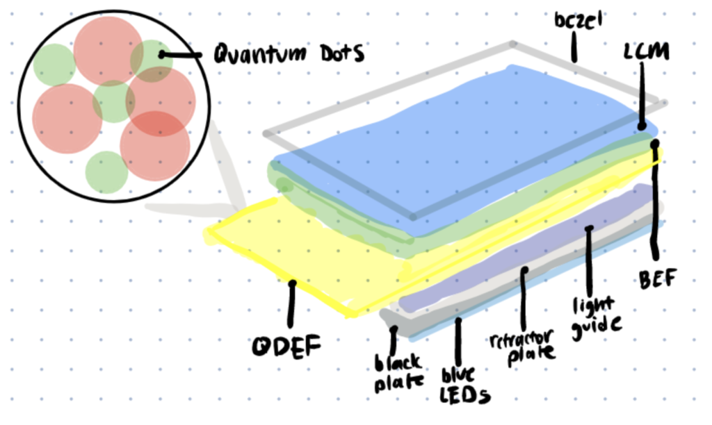
Several methods are proposed for using quantum dots to improve existing light-emitting diode (LED) design, including quantum dot LED (QLED) displays, and quantum dot white-LED (QWLED) displays. The main difference between QLEDs and other LED devices is that the light-emitting centers are cadmium selenide (CdSe), which provides a more reliable, energy-efficient, tunable color solution for display and lighting applications for high color saturation and stability.
QDs are photoluminescent and can produce monochromatic light naturally. These properties, coupled with their inherently good quantum yields, provides more saturated colors for higher efficacy and flexibility at a fraction of the energy cost compared to the traditional LED display.
Synthesis of Quantum Dots
Several routes have been used to synthesize QDs, but typical strategies for the synthesis of quantum dots include top-down and bottom-up approaches. Although top-down approaches are more affordable, scientists usually prefer to use bottom-up approaches, as they offer better control over chemical composition, morphology as well as better short-/long-range ordering of the QDs (Crista et al., 2020). A number of bottom-up approaches to generate QDs have been developed, but they may be broadly divided into wet-chemical and vapor-based methods, as summarized by Bera et al., 2010.
